Chemical warfare agent toxicology at the intersection of science and defense: a review of sarin and lewisite
DOI:
https://doi.org/10.55749/cds.v1i1.187Keywords:
Sarin and Lewisite, Chemical Warfare Agents, Toxicology, Chemical Terrorism, Medical CountermeasuresAbstract
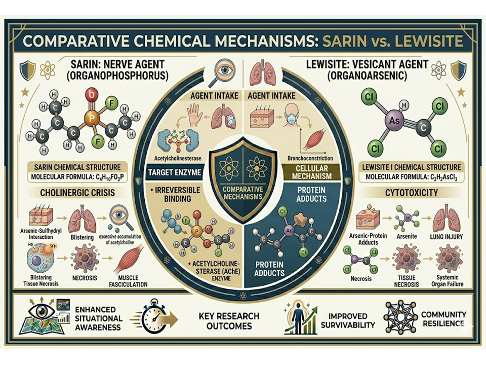
Chemical warfare agents (CWAs) remain a significant threat to military personnel, civilian populations, and public health systems due to their high toxicity and potential for mass casualties. This review compares the toxicological characteristics of sarin, a representative organophosphorus nerve agent, and Lewisite, an organoarsenic vesicant agent. Sarin exerts its toxicity primarily through irreversible inhibition of acetylcholinesterase, resulting in excessive accumulation of acetylcholine at cholinergic synapses and the development of acute cholinergic crisis characterized by miosis, bronchoconstriction, muscle fasciculations, seizures, respiratory failure, and death. In contrast, Lewisite induces cellular injury through the interaction of arsenic moieties with sulfhydryl-containing enzymes and proteins, disrupting essential metabolic pathways and leading to oxidative stress, tissue necrosis, vesication, respiratory injury, and systemic organ damage. The review discusses the physicochemical properties, toxicokinetics, biochemical mechanisms, clinical manifestations, diagnostic approaches, and medical management strategies associated with both agents. Historical incidents involving sarin and Lewisite are also examined to illustrate their operational significance and the challenges they pose to emergency preparedness and disaster response. Despite differences in their molecular targets and pathophysiological effects, both agents present substantial risks due to their rapid onset of toxicity and potential to overwhelm healthcare systems during large-scale exposures. A comprehensive understanding of these mechanisms is essential for improving risk assessment, clinical management, medical countermeasure development, and preparedness against chemical threats.
References
[1] Chai, P.R., Boyer, E.W., Al-Nahhas, H. and Erickson, T.B. 2017. Toxic chemical weapons of assassination and warfare: nerve agents VX and sarin. Toxicol. Commun. 1(1). 21-23. Doi: https://doi.org/10.1080/24734306.2017.1373503
[2] Abou-Donia, M.B., Siracuse, B., Gupta, N. and Sobel Sokol, A. 2016. Sarin (GB, O-isopropyl methylphosphonofluoridate) neurotoxicity: critical review. Crit. Rev. Toxicol. 46(10). 845-875. Doi: https://doi.org/10.1080/10408444.2016.1220916
[3] Costanzi, S., Machado, J.H. and Mitchell, M. 2018. Nerve agents: what they are, how they work, how to counter them. ACS Chem. Neurosci. 9(5). 873-885. Doi: https://doi.org/10.1021/acschemneuro.8b00148
[4] Gundert-Remy, U., Barth, H., Bürkle, A., Degen, G.H. and Landsiedel, R. 2015. Toxicology: a discipline in need of academic anchoring—the point of view of the German Society of Toxicology. Arch. Toxicol. 89(10). 1881-1893. Doi: https://doi.org/10.1007/s00204-015-1577-7
[5] Hayes, A.W. and Dixon, D. 2017. Cornerstones of toxicology. Toxicol. Pathol. 45(1). 57-63. Doi: https://doi.org/10.1177/0192623316675768
[6] Pognan, F., Beilmann, M., Boonen, H.C., Czich, A., Dear, G., Hewitt, P., Mow, T., Oinonen, T., Roth, A., Steger-Hartmann, T. and Valentin, J.P. 2023. The evolving role of investigative toxicology in the pharmaceutical industry. Nat. Rev. Drug. Discov. 22(4). 317-335. Doi: https://doi.org/10.1038/s41573-022-00633-x
[7] Shiek, S.S., Sajai, S.T. and Dsouza, H.S. 2023. Arsenic‐induced toxicity and the ameliorative role of antioxidants and natural compounds. J. Biochem. Mol. Toxicol. 37(3) 23281. Doi: https://doi.org/10.1002/jbt.23281
[8] Voros, C., Dias, J., Timperley, C.M., Nachon, F., Brown, R.C. and Baati, R. 2024. The risk associated with organophosphorus nerve agents: from their discovery to their unavoidable threat, current medical countermeasures and perspectives. Chem. Biol. Interact. 395. 110973. Doi: https://doi.org/10.1016/j.cbi.2024.110973
[9] Walczak-Nowicka, Ł.J. and Herbet, M. 2021. Acetylcholinesterase inhibitors in the treatment of neurodegenerative diseases and the role of acetylcholinesterase in their pathogenesis. Int. J. Mol. Sci. 22(17). 9290. Doi: https://doi.org/10.3390/ijms22179290
[10] Lagacé, F., Schmitt, A., Martrille, L., Benassi, J. and Adalian, P. 2020. Using histomorphometry for human and nonhuman distinction: A test of four methods on fresh and archaeological fragmented bones. Forensic. Sci. Int. 313. 110369. Doi: https://doi.org/10.1016/j.forsciint.2020.110369
[11] Bernardo, L.B., Borges, C.V., Buitrago, P.A., Kuča, K., Cavalcante, S.F., Sousa, R.B., Lima, A.L. and Kitagawa, D.A. 2024. Synthesis and in vitro assessment of the reactivation profile of clinically available oximes on the acetylcholinesterase model inhibited by A-230 nerve agent surrogate. Arch. Toxicol. 98(10). 3397-3407. Doi: https:// doi.org/10.26434/chemrxiv-2024-xkp1w
[12] Meyer, C. and Thippeswamy, T. 2025. Organophosphate Chemical Nerve Agents, Oxidative Stress, and NADPH Oxidase Inhibitors: An Overview. Int. J. Mol. Sci. 26(19). 9313. Doi: https://doi.org/10.3390/ijms26199313
[13] Cao, C., Wang, L., Zhang, J., Liu, Z., Li, M., Xie, S., Chen, G. and Xu, X. 2022. Neuroligin-1 plays an important role in methamphetamine-induced hippocampal synaptic plasticity. Toxicol. Lett. 361. 1-9. Doi: https://doi.org/10.1016/j.toxlet.2022.03.007
[14] Huber, C. 2019. A war of frustration: Saddam Hussein’s use of nerve gas on civilians at Halabja (1988) and the American response. Senior Honors. 6(03) .2022. Doi: https://commons.lib.jmu.edu/honors201019/683
[15] Etemad, L., Moshiri, M. and Balali-Mood, M. 2018. Delayed Complications and long-term management of sulfur mustard poisoning: a narrative review of recent advances by iranian researchers part іі: clinical management and therapy. Iran. J. Med. Sci. 43(3). 235.
[16] Romero Martínez, M.A., Ramos Alonso, E., López Muñoz, F., Ríos, C.D.L., Egea, J., Gil Martín, E., Pita Pita, R., Torrado Durán, J.J., Serrano López, D.R. and Juberias, A., 2021. Toxicology of Blister Agents: Is Melatonin a Potential Therapeutic Option?. Diseases. 9(2). 27. Doi: https://doi.org/10.3390/diseases9020027
[17] Li, C., Srivastava, R.K. and Athar, M. 2016. Biological and environmental hazards associated with exposure to chemical warfare agents: arsenicals. Annals Ann. N.Y. Acad. Sci. 1378(1). 143-157. Doi: https://doi.org/10.1111/nyas.13214
[18] Li, C., Srivastava, R.K. and Athar, M. 2016. Biological and environmental hazards associated with exposure to chemical warfare agents: arsenicals. Ann. N. Y. Acad. Sci. 1378(1). 143-157. Doi: https://doi.org/10.1111/nyas.13214
[19] Muzaffar, S., Khan, J., Srivastava, R., Gorbatyuk, M.S. and Athar, M. 2023. Mechanistic understanding of the toxic effects of arsenic and warfare arsenicals on human health and environment. Cell Biol. Toxicol. 39(1). 85-110. Doi: https://doi.org/10.1007/s10565-022-09710-8
[20] Sugiyama, A., Matsuoka, T., Sakamune, K., Akita, T., Makita, R., Kimura, S., Kuroiwa, Y., Nagao, M. and Tanaka, J. 2020. The Tokyo subway sarin attack has long-term effects on survivors: A 10-year study started 5 years after the terrorist incident. PloS one. 15(6). 0234967. Doi: https://doi.org/10.1371/journal.pone.0234967
[21] Socher, M., Romanek, K., Zilker, T., Fromme, H. and Wildner, M. 2025. Assaults with Highly Toxic Substances in Public Spaces: Preparedness in Lessons Learnt from Precedent Cases. Doi: https://doi.org/10.5772/intechopen.1011887
[22] Baker, D.J. and Sedgwick, E.M. 1996. Single fibre electromyographic changes in man after organophosphate exposure. Hum. Exp. Toxicol. 15(5). 369-375. Doi: https://doi.org/10.1177/096032719601500501
[23] Makhaeva, G.F., Rudakova, E.V., Sigolaeva, L.V., Kurochkin, I.N. and Richardson, R.J. 2016. Neuropathy target esterase in mouse whole blood as a biomarker of exposure to neuropathic organophosphorus compounds. J. Appl. Toxicol. 36(11). 1468-1475. Doi: https://doi.org/10.1002/jat.3305
[24] Srivastava, R.K., Muzaffar, S., Khan, J., Bansal, M., Agarwal, A. and Athar, M. 2025. Common molecular profile of multiple structurally distinct warfare arsenicals in causing cutaneous chemical vesicant injury. Sci. Rep. 15(1). 6505. Doi: https://doi.org/10.1038/s41598-024-83513-1
[25] Milatović, D., Jokanović, M. and Gupta, R.C., 2009. Handbook of toxicology of chemical warfare agents.
[26] Gorbatyuk, M., Sinha, N.R., Kumar, R., Zhylkibayev, A., Athar, M., Patrick, M. and Mohan, R.R. 2025. Current progress in research on ocular injury caused by exposure to vesicants. Prog. Retin. Eye. Res. 109. 101413. Doi: https://doi.org/10.1016/j.preteyeres.2025.101413
[27] Nair, A., Yadav, P., Behl, A., Sharma, R.K., Kulshrestha, S., Butola, B.S. and Sharma, N., 2021. Toxic blister agents: Chemistry, mode of their action and effective treatment strategies. Chem. Biol. Interact. 350. 109654. Doi: https://doi.org/10.1016/j.cbi.2021.109654
[28] Allgardsson, A., Berg, L., Akfur, C., Hörnberg, A., Worek, F., Linusson, A. and Ekström, F.J. 2016. Structure of a prereaction complex between the nerve agent sarin, its biological target acetylcholinesterase, and the antidote HI-6. PNAS. 113(20). 5514-5519. Doi: https://doi.org/10.1073/pnas.1523362113
[29] Abou-Donia, M.B., Siracuse, B., Gupta, N. and Sobel Sokol, A. 2016. Sarin (GB, O-isopropyl methylphosphonofluoridate) neurotoxicity: critical review. Crit. Rev. Toxicol. 46(10). 845-875. Doi: https://doi.org/10.1080/10408444.2016.1220916
[30] Zhuang, Q., Franjesevic, A.J., Corrigan, T.S., Coldren, W.H., Dicken, R., Sillart, S., DeYong, A., Yoshino, N., Smith, J., Fabry, S. and Fitzpatrick, K. 2018. Demonstration of in vitro resurrection of aged acetylcholinesterase after exposure to organophosphorus chemical nerve agents. J. Med. Chem. 61(16). 7034-7042. Doi: https://doi.org/10.1021/acs.jmedchem.7b01620
[31] Aroniadou-Anderjaska, V., Apland, J.P., Figueiredo, T.H., Furtado, M.D.A. and Braga, M.F. 2020. Acetylcholinesterase inhibitors (nerve agents) as weapons of mass destruction: History, mechanisms of action, and medical countermeasures. Neuropharmacology. 181. 108298. Doi: https://doi.org/10.1016/j.neuropharm.2020.108298
[32] Harper, L.K. and Bayse, C.A., 2015. Modeling the chelation of As (III) in lewisite by dithiols using density functional theory and solvent-assisted proton exchange. J. Inorg. Biochem. 153. 60-67. Doi: https://doi.org/10.1016/j.jinorgbio.2015.10.004
[33] Nurchi, V.M., Buha Djordjevic, A., Crisponi, G., Alexander, J., Bjørklund, G. and Aaseth, J. 2020. Arsenic toxicity: molecular targets and therapeutic agents. Biomolecules. 10(2). 235. Doi: https://doi.org/10.3390/biom10020235
[34] Lyczko, M., Lyczko, K., Majkowska-Pilip, A. and Bilewicz, A. 2019. 1, 2-Benzenedithiol and Toluene-3, 4-dithiol Arsenic (III) Complexes—Synthesis, Structure, Spectroscopic Characterization and Toxicological Studies. Molecules. 24(21). 3865. Doi: https://doi.org/10.3390/molecules24213865
[35] Yu, F.L., Zhang, M.Y., Song, G.W., Ning, Y.S., Wu, X. and Gao, Y., 2025. The effect of arsenic on mitochondrial fatty acid metabolism via inhibition of carnitine palmitoyltransferase 1B and choline kinase beta in C2C12 cells. Plos one. 20(5). e0320557. Doi: https://doi.org/10.1371/journal.pone.0320557
[36] Turkington, R.E., Hukriede, N.A., Ho, J., Jayasundara, N. and Sanders, A.P. 2025. Metal mechanisms of mitochondrial toxicity: recent review of arsenic, cadmium, and lead-induced nephrotoxicity. Environ. Sci. Pollut. Res. 32(24). 14439-14451. Doi: https://doi.org/10.1007/s11356-025-36538-6
[37] Jett, D.A., Sibrizzi, C.A., Blain, R.B., Hartman, P.A., Lein, P.J., Taylor, K.W. and Rooney, A.A. 2020. A national toxicology program systematic review of the evidence for long-term effects after acute exposure to sarin nerve agent. Crit. Rev. Toxicol. 50(6). 474-490. Doi: https://doi.org/10.1080/10408444.2020.1787330
[38] Lee, T.J. and Borgman, M.A. 2022. Toxic Inhalations. In Management of Chest Trauma: A Practical Guide. 367-385. Cham: Springer International Publishing. Doi: https://doi.org/10.1007/978-3-031-06959-8_31
[39] Hatfill, S.J. 2019. Chemical warfare: nerve agents. J. Am. Phys. Surg. 24(1). 19-24.
[40] Howard Jr, J.F. 2018. Toxic neuromuscular transmission disorders. In Myasthenia gravis and related disorders. 275-298. Cham: Springer International Publishing. Doi: https://doi.org/10.1007/978-3-319-73585-6_17
[41] Andrew, P.M. and Lein, P.J. 2021. Neuroinflammation as a therapeutic target for mitigating the long-term consequences of acute organophosphate intoxication. Front. Pharmacol. 12. 674325. Doi: https://doi.org/10.3389/fphar.2021.674325
[42] John, H. and Thiermann, H. 2021. Poisoning by organophosphorus nerve agents and pesticides: an overview of the principle strategies and current progress of mass spectrometry-based procedures for verification. J. Mass Spectrom. Adv. Clin. Lab. 19. 20-31. Doi: https://doi.org/10.1016/j.jmsacl.2021.01.002
[43] Norrrahim, M.N.F., Razak, M.A.I.A., Shah, N.A.A., Kasim, H., Yusoff, W.Y.W., Halim, N.A., Nor, S.A.M., Jamal, S.H., Ong, K.K., Yunus, W.M.Z.W. and Knight, V.F. 2020. Recent developments on oximes to improve the blood brain barrier penetration for the treatment of organophosphorus poisoning: a review. RSC Adv. 10(8). 4465-4489. Doi: https://doi.org/10.1039/C9RA08599H
[44] Labaška, M., Gál, M., Mackuľak, T., Švorec, J., Kučera, J., Helenin, J., Svitková, V. and Ryba, J. 2024. Neutralizing the threat: A comprehensive review of chemical warfare agent decontamination strategies. J. Environ. Chem. Eng. 12(6). 114243. Doi: https://doi.org/10.1016/j.jece.2024.114243
[45] Dai, X. and Shen, L. 2022. Advances and trends in omics technology development. Front. Med. 9. 911861. Doi: https://doi.org/10.3389/fmed.2022.911861
[46] Mohamed, R.A., Ong, K.K., M. Kasim, N.A., A. Halim, N., M. Noor, S.A., Knight, V.F., Ab. Rahman, N.N. and W. Yunus, W.M.Z. 2021. Transitioning from oxime to the next potential organophosphorus poisoning therapy using enzymes. J. Chem. 2021(1). 7319588. Doi: https://doi.org/10.1155/2021/7319588
[47] Balali-Mood, M., Eizadi-Mood, N., Hassanian-Moghaddam, H., Etemad, L., Moshiri, M., Vahabzadeh, M. and Sadeghi, M. 2025. Recent advances in the clinical management of intoxication by five heavy metals: Mercury, lead, chromium, cadmium and arsenic. Heliyon. 11(4). Doi: https://doi.org/10.1016/j.heliyon.2025.e42696
[48] Bjørklund, G., Oliinyk, P., Lysiuk, R., Rahaman, M.S., Antonyak, H., Lozynska, I., Lenchyk, L. and Peana, M. 2020. Arsenic intoxication: general aspects and chelating agents. Arch. Toxicol. 94(6), 1879-1897. Doi: https://doi.org/10.1007/s00204-020-02739-w
[49] Kosnett, M.J. 2013. The role of chelation in the treatment of arsenic and mercury poisoning. J. Med. Toxicol. 9(4). 347-354. Doi: https://doi.org/10.1007/s13181-013-0344-5
[50] Calvano, C.J., Enzenauer, R.W. and Johnson, A.J. eds. 2019. Ophthalmology in Military and Civilian Casualty Care. No. 168443. Springer. Doi: https://doi.org/10.1007/978-3-030-14437-1
[51] Singh, A.V., Bhardwaj, P., Laux, P., Pradeep, P., Busse, M., Luch, A., Hirose, A., Osgood, C.J. and Stacey, M.W. 2024. AI and ML-based risk assessment of chemicals: predicting carcinogenic risk from chemical-induced genomic instability. Front. Toxicol. 6. 1461587. Doi: https://doi.org/10.3389/ftox.2024.1461587
[52] Hartung, T. 2023. Artificial intelligence as the new frontier in chemical risk assessment. Front. Artif. Intell. 6, p.1269932. Doi: https://doi.org/10.3389/frai.2023.1269932
Downloads
Published
How to Cite
Issue
Section
License
Copyright (c) 2026 CBRNE Defense Studies

This work is licensed under a Creative Commons Attribution-ShareAlike 4.0 International License.